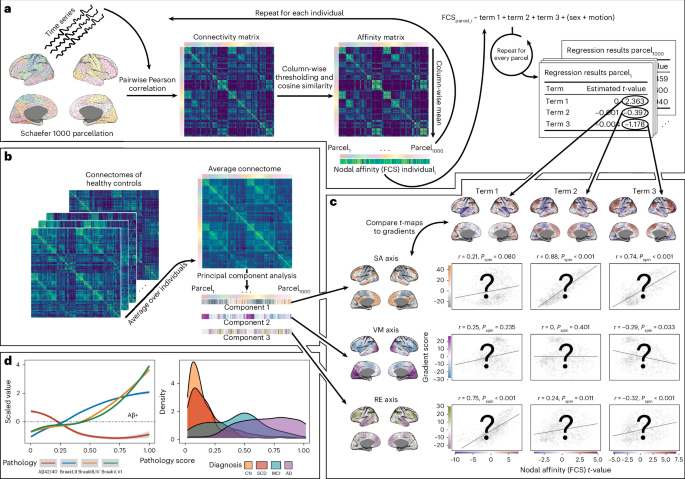
Aging and Alzheimer’s disease (AD) are accompanied by alterations to large-scale communication patterns in the brain, which can be tracked in vivo using functional connectivity. The location, direction and relevance of these changes remain widely debated, although they are rarely studied in the context of whole-cortex communication dynamics. Here, in two independent cohorts (BioFINDER-2, N = 973; Alzheimer’s Disease Neuroimaging Initiative, N = 129), we show that functional connectivity changes associated with aging and AD are strongly aligned with separate fundamental axes of hierarchical brain communication.
Early accumulation of AD pathology and subsequent cognitive decline are both linked to functional change along the sensory–association axis. Meanwhile, age-related functional changes occur along the representation–executive axis consistently throughout the adult lifespan. These findings together suggest that both AD and aging alter major but orthogonal functional pathways in the brain.
More broadly, our findings position whole-brain connectivity dynamics as a unifying framework for interpreting functional changes across the adult lifespan. Aging and Alzheimer’s disease (AD) are both associated with changes in functional brain networks—groups of regions that typically communicate with one another and support cognition. However, the precise nature of these changes—their spatio-temporal dynamics and whether they reflect compensatory adaptations, pathological disruptions or both—remains a topic of active debate1,2.
Answering these questions is not only important for understanding disease biology but could enable tailored clinical strategies that address age-independent mechanisms of AD. AD is the leading cause of dementia and is characterized by pathological accumulation of amyloid-β and tau proteins. These proteins spread through the brain in characteristic patterns3,4, leading to neurodegeneration and cognitive decline.
Functional connectivity (FC), the temporal synchrony of activity between brain regions, may be a mechanism facilitating the spread of pathology, a system affected by pathology or an interplay of both5. However, normal or healthy aging is also associated with altered FC6,7,8. These age- and AD-related alterations have been proposed both as mechanisms to sustain cognitive function, as the brain is challenged by stressors, and as manifestations of pathological network breakdown2,9,10,11,12.
FC changes in aging and AD have commonly been studied through two approaches. One approach focuses on regional connectivity changes, such as increases or decreases within or between specific regions or networks, or between regions of interest and the rest of the brain13,14. Although some consistent effects have been reported, such as reduced FC within the default mode network (DMN) in healthy aging7, the overall picture is more complex.
Extract — continue reading at the source.


