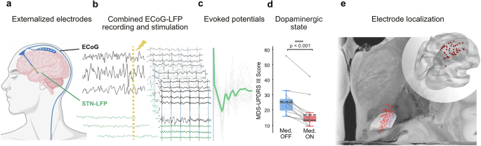
The cortico-subthalamic hyperdirect pathway provides a rapid route for cortical influence over basal ganglia processing and is a key target of deep brain stimulation (DBS) in Parkinson’s disease (PD). However, to what degree dopamine modulates signal transmission along this pathway in humans remains unclear. Using bidirectional cortical and subthalamic electrical stimulation while invasively recording evoked responses in 15 PD patients undergoing DBS surgery, we examined cortico-subthalamic communication across dopaminergic states.
Cortical stimulation evoked a short-latency subthalamic response consistent with monosynaptic hyperdirect activation that was preserved across medication conditions. In contrast, longer-latency subthalamic responses showed significant dopaminergic modulation, while cortical responses to subthalamic stimulation remained unchanged. These results reveal a temporal dissociation within the cortex–basal ganglia network, whereby fast cortical transmission is unaffected by dopamine, which in turn amplifies slower network level communication to the subthalamic nucleus.
This separation of fast and delayed components provides a mechanistic framework for understanding how dopamine shapes information flow in the cortex–basal ganglia system. Parkinson’s disease (PD) is the most rapidly expanding neurodegenerative condition1,2, characterized by the progressive degeneration of dopaminergic neurons within the substantia nigra3. Dopamine replacement therapy with levodopa, together with high-frequency deep brain stimulation (DBS) of the subthalamic nucleus (STN), remain the most effective interventions for alleviating PD-related motor symptoms4,5.
Yet despite their widespread clinical use, the neural circuit mechanisms through which these treatments exert their therapeutic effects are still not fully understood6. One of the proposed target circuits of STN-DBS is the monosynaptic cortico-subthalamic hyperdirect pathway, which has been implicated in rapid movement cancellation, in the emergence of pathological synchronization, and as a key substrate through which STN-DBS may influence network dynamics7,8,9,10. Although computational models and animal experiments have highlighted the importance of this pathway11,12,13, direct causal evidence for how dopaminergic state shapes hyperdirect STN excitation in humans remains scarce9,14.
This pathway can be probed electrophysiologically using evoked responses following electrical stimulation, which provide a time-resolved measure of signal propagation along cortico-subthalamic circuits. Our main objective is to test whether dopaminergic state differentially modulates fast versus delayed cortico-subthalamic evoked responses in humans. In this context, short-latency responses are thought to reflect fast, likely monosynaptic transmission along the hyperdirect pathway, whereas longer-latency responses are more likely to reflect polysynaptic network activity.
Prior human studies have relied primarily on stimulation of the STN to measure antidromic activation with electrocorticography (ECoG), electroencephalography (EEG) or magnetoencephalography (MEG) recordings15,16,17,18,19. However, it remains unclear whether this cortical neural population activity behaves similarly to orthodromic activation and, if so, to what extent this activity can be modulated by dopamine. To address this gap, we implemented direct electrical stimulation of motor cortex delivered through externalized ECoG electrodes, during simultaneous STN local field potential (LFP) recordings in PD patients undergoing DBS implantation.
Extract — continue reading at the source.


