Familial Parkinson’s disease (PD) and vascular parkinsonism (VP) present overlapping features and may co-exist. To investigate whether PD and VP may share a potential pathogenic link and to what extent white matter hyperintensities may influence PD phenotype, we used the modified Scheltens scale and presented a descriptive and exploratory analysis of the classic neuroradiological features of cerebral small vessel disease (cSVD) in the axial T2-FLAIR MRI sequences in a cohort of 104 familial PD and PD prodromal patients and 48 age-matched controls from the PPMI publicly available database. We next performed whole exome sequencing to examine the protein coding variability in the main PD-causing and risk genes (VPS35, DJ1, PINK1, ATP13A2, PRKN, SNCA, LRRK2, GBA, MAPT, LAMP3, STK39) in a cohort of 96 patients with familial cSVD and 243 elderly healthy individuals (HEX database).
In this cohort, patients with familial and prodromal PD present a moderate burden of superficial frontal white matter hyperintensities (p-value = 3.46e-06, Bonferroni-corrected), linked to a mild reduction of motor and cognitive function and an increased LRRK2 p.G2019S and p.R1441C variant penetrance, and bilateral basal ganglia periventricular enlarged spaces (p-value = 2.64e-03, Bonferroni-corrected). Moreover, one-third of familial PD patients displayed a burden of lacunar thalamic strokes (p-value = 0.058), associated with a moderate hypokinetic-rigid syndrome. Finally, we report no known pathogenic coding variant in the main PD causative genes and risk factors in a cohort of 96 early-onset cSVD Caucasian patients.
Our study adds to the understanding of potential cSVD hallmarks within this familial LRRK2, GBA and SNCA PD-PD prodromal cohort. Vascular parkinsonism is often caused by sporadic, severe, diffuse progressive cerebral small vessel disease (cSVD). Other less common causes of VP include Mendelian cSVDs, such as Cerebral Autosomal Dominant Arteriopathy with Subcortical Infarcts and Leukoencephalopathy (CADASIL), Cerebral Autosomal Recessive Arteriopathy with Subcortical Infarcts and Leukoencephalopathy (CARASIL), and retinal vasculopathy with cerebral leukodystrophy (RVCL).
More rarely, Mendelian leukodystrophies such as hereditary diffuse leukoencephalopathy with spheroids (HDLS) leading to a reduction of thalamo-cortical drive may also cause VP2,3 (https://www.omim.org/) (Table S2). However, despite the comprehensive body of both clinical and experimental evidence describing white matter macro- and microstructural changes in PD patients and correlating these to the progression of diverse clinical PD phenotypes4–5, LRRK2 p.G2019S variant leading to abnormalities in arterial cerebral blood flow in mouse experimental models6, a potential common pathogenic ground between familial PD and cSVD has not been extensively and systematically investigated. Moreover, although brain vascular lesions have been described in almost 25% of patients with idiopathic PD7, their contribution to the clinical phenotypic spectrum remains to be unveiled.
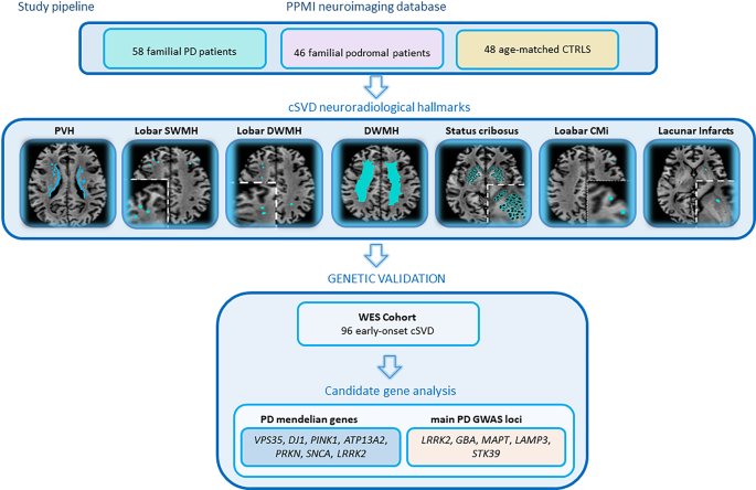
Therefore, we used a modified Scheltens scale and screened the classical cSVD neuroradiological biomarkers (periventricular hyperintensities, lobar superficial white matter hyperintensities, lobar deep white matter hyperintensities, status cribrosus, lobar cortical small-microinfarcts, lacunar infarcts8–9, Table S3) in T2-FLAIR MRI sequences of 58 familial PD and 46 familial PD prodromal patients and 48 age-matched controls from the PPMI publicly available database10(Table 1, Table S4). Next, we performed exome sequencing on a cohort of 96 familial cSVD Caucasian patients from the US to investigate in this cohort protein coding variability in the most common PD Mendelian genes (VPS35, DJ1, PINK1, ATP13A2, PRKN, SNCA, LRRK2) and risk factors (LRRK2, GBA, MAPT, LAMP3, STK39) (https://www.omim.org/) (Fig. 1). Fifty-eight familial Parkinson’s disease patients (females = 22, mean age at onset 60 years [range: 32–82]), 46 familial prodromal PD patients (females = 27, mean age at of onset 63 years [range: 34–77]) and 48 age-matched healthy controls (females = 17, mean age at onset 61 years [range: 32–81]) were obtained from the Parkinson Progression Marker Initiative (PPMI) database (Table 1, Table S4).
We downloaded data on PD patients from the PPMI database in December 2021. The PPMI study is well described at www.ppmi-info.org10. Briefly, PPMI is an observational, comprehensive multi-center study designed to identify biomarkers of PD with the goal of improving evaluation of disease-modifying therapeutics11.
Extract — continue reading at the source.


