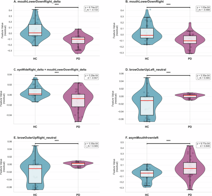
Reduced facial expressivity (hypomimia) is an early motor feature of Parkinson’s disease (PD), yet its assessment still relies on subjective, coarse-grained clinical scales. Using ExpressionTracker, a consumer-grade application built on Apple’s ARKit framework, we derived 261 facial expression features during standardised cued expression tasks in 34 people with PD and 36 healthy controls (HC). Beyond the expected reduction in movement amplitude, PD was characterised by increased facial asymmetry and attenuated activity of ipsilateral mouth and eye regions.
Interaction and asymmetry features were prominent among the between-group differences and explained 38 to 42 percent of the variance in clinician-rated hypomimia. For diagnostic classification (PD versus HC) under nested cross-validation, the best model (gradient boosting machine, GBM) reached an area under the receiver operating characteristic curve (AUC) of 0.834 (95% CI 0.733 to 0.923), followed by XGBoost (0.810, 95% CI 0.702 to 0.906) and light gradient boosting machine (LightGBM; 0.804, 95% CI 0.697 to 0.899). Engineered ARKit features outperformed an amplitude-only baseline (maximum AUC 0.75, 95% CI 0.63 to 0.86) and a demographic confounder-only baseline using age and sex (maximum AUC 0.68, 95% CI 0.55 to 0.80).
These results indicate that automated facial blendshape analysis captures multidimensional motor dysfunction beyond amplitude reduction alone, positioning ExpressionTracker as a candidate digital biomarker for community screening, disease monitoring, and drug-efficacy assessment in future clinical trials. Hypomimia, or reduced facial expressivity, is one of the earliest motor hallmarks of Parkinson’s disease (PD) and affects both spontaneous and voluntary facial movements1. Hypomimia can precede the clinical diagnosis of PD by up to ten years in individuals with idiopathic REM sleep behaviour disorder2,3.
Current assessment of hypomimia relies on semiquantitative clinical judgement using item 3.2 (facial expression) of the Movement Disorder Society Unified Parkinson’s Disease Rating Scale Part III (MDS-UPDRS III), which grades hypomimia from reduced blink frequency, diminished perioral movement including spontaneous smiling, and parted lips at rest4. Although valuable and extensively validated in routine practice, the MDS-UPDRS III was not developed for early-stage PD and remains subject to substantial interrater variability and to floor and ceiling effects5,6,7. Moreover, it is a nonlinear ordinal scale rather than an interval measure, so that differences between adjacent scores are not necessarily equivalent.
As disease-modifying therapies advance toward clinical trials yet repeatedly fail to demonstrate efficacy, there is a pressing need to characterise and quantify all phenomenological aspects of PD, including hypomimia, in an objective and standardised manner. Cumulative evidence supports computer vision for automated hypomimia detection, but the approaches applied to date have been heterogeneous. A recent systematic review groups them into method families that span task-based paradigms such as reading aloud, emotion-elicitation tasks such as posed smiling, anger, or disgust, and data-mining of spontaneous expressions extracted from internet images and videos8,9,10,11,12.
Within these families, methods range from facial-landmark and distance metrics, through action-unit pipelines such as OpenFace coupled with logistic regression, to deep-learning classifiers trained on video, often with data augmentation to offset small samples, and to studies of spontaneous expressivity13,14,15. Two limitations recur across these families. First, many paradigms rely predominantly on visual emotion-recognition cues without accounting for the impaired emotion recognition frequently observed in PD, which can confound measurements of voluntary facial expressiveness16,17,18,19.
Extract — continue reading at the source.


