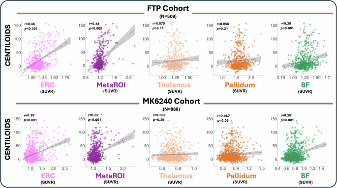
The basal forebrain (BF) is a site of early tau pathology deposition, and BF cholinergic neurons selectively degenerate in patients with Alzheimer’s disease (AD). PET imaging of AD pathology has helped to establish the relationships between early tau in the medial temporal lobe (MTL), amyloid (Aβ) pathology, brain structure and cognition, but it is not clear if PET imaging can be used to similarly characterize early tau pathology in the BF. Here we aim to test the feasibility of measuring tau pathology in the BF in unimpaired older adults with two PET tracers, [18F]flortaucipir (FTP) and [18F]MK6240, that have unique properties including distinct off-target signal (OTS) patterns. 1397 unimpaired older adult participants with either FTP (n = 509; Berkeley Aging Cohort Study, ADNI) or MK6240 (n = 888; POINTER Imaging study) scans were included and used to quantify tau-PET signal in three ROI categories: the BF, typical temporal tau-PET ROIs (entorhinal cortex [ERC], temporal MetaROI) and OTS ROIs that are adjacent to the BF (thalamus, pallidum).
BF and temporal tau burden were associated with global Aβ burden, but OTS was not. BF and MTL tau showed similar associations with age, BF volume, hippocampal volume and multi-domain cognition. Associations were consistent in both FTP and MK6240 cohorts.
MTL tau was negatively correlated with BF volume in whole-brain morphometry analyses. Our findings suggest that it is feasible to use PET to measure tau signal in the BF in unimpaired older adults. The basal forebrain (BF) region is located ventral to the anterior commissure and contains a collection of nuclei, including the nucleus basalis of Meynert (nbM) which is vulnerable to Alzheimer’s disease (AD) [1,2,3].
The cholinergic neurons of the nbM are known to degenerate in AD and this observation led to the cholinergic hypothesis of AD [4] and the development of cholinesterase inhibitors to treat AD symptoms [5, 6]. Critically, neuropathology studies show that the BF is a site of early tau pathology deposition [7], but the impact of this early tau pathology is not well-understood, especially when compared to the more established relationship between early medial temporal lobe (MTL) tau and episodic memory performance [8, 9]. The link between MTL tau and downstream effects on brain structure and episodic memory in preclinical (asymptomatic) AD has been shown over the past decade in tau-PET studies that enabled in-vivo measurements of regional tau in cognitively normal older adults [10,11,12,13].
Tau-PET measurements of pathology in the BF would allow us to investigate downstream effects of BF tau and compare them to MTL tau. Quantifying tau-PET in the BF has several potential challenges including the small size of the region, its heterogenous components (nbM, diagonal band of Broca, septal nuclei) and its proximity to regions of known off-target signal (OTS) for some tau-PET tracers [14,15,16,17]. Despite these potential challenges, we set out to determine the feasibility of measuring early tau pathology with tau-PET in the BF.
To do this, we used two large cohorts of cognitively healthy older adults imaged either with [18F]Flortaucipir (FTP) or [18F]MK6240. These two tracers have different tau affinities and different patterns of OTS, which allowed us to also explore whether feasibility of measuring BF tau-PET signal differed by tracer. Using a previously developed, functionally-defined BF ROI [18], we calculated cross-sectional BF tau-PET signal and BF gray matter (GM) volume for each participant.
Extract — continue reading at the source.


