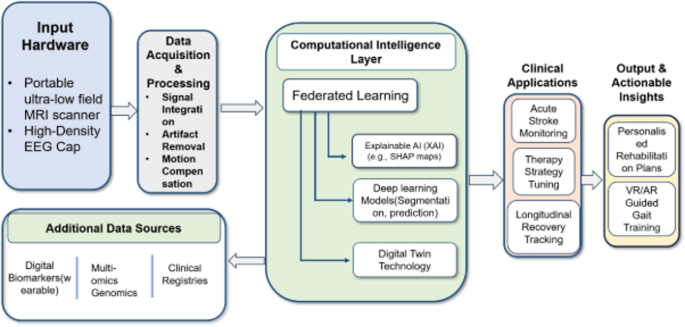
The occurrence of stroke continues to be one of the leading causes of disability in the world today, stressing the importance of a correct diagnosis and rehabilitation strategy that takes into account the dynamic process of brain reorganization that follows such an event. While electroencephalography (EEG) is used to study neuronal activity on the millisecond scale, magnetic resonance imaging (MRI) provides high-resolution structural and functional images. While it remains that the choice for the acute setting as an imaging modality is MRI/CT to inform treatment decisions, it can increasingly be argued that linking MRI to EEG becomes more valuable for prognosis, intensity assessment, and rehabilitation at this level of care.
In this narrative review, we discuss recent advancements in EEG-MRI signal processing for ischemic and hemorrhagic stroke, with particular attention to preprocessing, artifact removal, feature extraction, multimodal data fusion, and machine learning methods. In addition to presenting the existing methods for stroke-related multimodal applications, we highlight current challenges for translating these techniques from research to clinical practice, which include the limited availability of publicly available multimodal data, a lack of longitudinal studies covering acute to chronic phases of recovery, inconsistency in standardization of acquisition and preprocessing protocols, and neglecting applications related to rehabilitation. It is important to note that this review provides an overview of how emerging technologies can address the problems mentioned above.
This review highlights developments in neuroimaging, signal processing, artificial intelligence, and rehabilitation science, and discusses the potential applications of these techniques in stroke treatment. Stroke is an acute cerebrovascular event characterized by sudden interruption of blood flow in the brain, leading to subsequent tissue damage resulting (depending on the cause) in ischemic or hemorrhagic injury and typically to an acute deficit of function. The epidemiology is significant, with stroke being the third leading cause of disability-adjusted life years (DALYs) globally and with incidence and mortality for stroke growing globally between 1990 and 2021 even though the age-adjusted rates have declined1.
There are two major types of strokes: ischemic (thrombo-embolic arterial occlusion that leads to focal hypoperfusion of brain parenchyma) and hemorrhagic (rupture of a brain vessel and intraparenchymal hemorrhage), respectively. In both subtypes and especially within the “ischemic penumbra”, or that severely hypoperfused but not yet infarcted portion of the brain immediately adjacent to the definitively infarcted zone, neuronal injury propagates over a relatively short, individual-specific therapeutic window, suggesting a necessity for acute identification and hyperacute treatment to preserve at-risk brain and maximize functional recovery2. Neuroimaging has played a vital role in the current management workflow for triaging patients, estimating prognosis, and following up on rehabilitation efforts, in fact replacing CT as the primary neuroimaging modality for structural evaluation to a large extent due to MRI’s better soft-tissue contrast.
In addition, certain MRI sequences provide non-structural, biophysical data, such as diffusion or perfusion imaging parameters, that are valuable3. Another specialized technique, perfusion-weighted imaging (PWI), estimates regional cerebral blood flow and transit-time kinetics to localize hypoperfused tissue, including both the stroke core and salvageable penumbra2. In contrast, diffusion-weighted imaging (DWI) and diffusion tensor imaging (DTI) evaluate white-matter integrity by characterizing the direction and extent of water diffusion to characterize the microstructure of fiber tracts such as the corticospinal tract4.
Functional MRI (fMRI), which relies on the blood-oxygen-level-dependent (BOLD) signal to study the connectivity and dynamics of neural networks at millimeter spatial resolution, can also map the effects of stroke3. The sensitivity of each technique to detect changes after stroke will depend on the appropriate spatial and temporal resolution. Electroencephalography (EEG), however, measures directly brain electrical activity from the scalp surface and reflects the synchronous postsynaptic potentials of populations of cortical neurons on the timescale of milliseconds5.
Extract — continue reading at the source.


