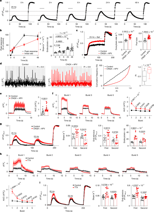
The neural plasticity that underlies adaptation and learning requires countervailing homeostatic mechanisms to maintain the stability and responsiveness of neural circuits. Previous work has described several forms of synaptic homeostasis1, but the mechanisms that detect changes in activity and transduce these into altered neurotransmission have remained poorly understood. Here we show that high-frequency stimulation alters the subsequent properties of neurotransmitter release to confer a presynaptic form of homeostasis, independent of postsynaptic receptors.
The mechanism involves vesicular SNARE protein (v-SNARE) VAMP7 (vesicle-associated membrane protein 7), which reduces neurotransmitter release by inhibiting the entry of Ca2+ specifically through P/Q-type presynaptic Ca2+ channels. Previous work has implicated target SNARE proteins (t-SNAREs) in the inhibition of Ca2+ channels2, but without clear physiological rationale. We show that the activity-dependent regulation of release mediated by VAMP7 requires the t-SNARE syntaxin.
Although VAMP7 acts at the plasma membrane to inhibit P/Q-type channels, it localizes predominantly to a subset of synaptic vesicles made by adaptor protein-3 (AP-3) that respond specifically to high-frequency stimulation3,4. The localization of VAMP7 to AP-3-derived synaptic vesicles thus provides a mechanism for the activity-dependent delivery of VAMP7 to the plasma membrane, where it associates with syntaxin to inhibit P/Q-type Ca2+ channels and transmitter release. This homeostatic form of metaplasticity operates at multiple excitatory synapses, and in its absence, the fraction of FOS+ neurons increases several fold, revealing a general property of excitatory neurotransmission that might also maintain the sparsity of neural connections.
Homeostatic synaptic plasticity can involve pre- or postsynaptic mechanisms. In response to a global change in activity, mammalian central neurons alter the expression of glutamate receptors1,5,6,7. By contrast, a presynaptic form of homeostasis occurs at the neuromuscular junction.
First established in Drosophila melanogaster, this involves increased release of glutamate in response to reduced postsynaptic receptor function8,9. An effect of activity on neurotransmitter release has also been reported at central synapses10,11,12,13,14,15,16,17,18. Although previous work has identified several elements that are required for the different forms of homeostatic plasticity, the mechanisms involved remain poorly understood.
To determine the time course over which presynaptic homeostatic plasticity develops, we suppressed network activity by treating primary mouse hippocampal cultures, which contain almost entirely glutamatergic neurons (Extended Data Fig. 1a,b), with the glutamate receptor antagonists 6-cyano-7-nitroquinoxaline-2,3-dione (CNQX) and 3-(2-carboxypiperazin-4-yl)propyl-1-phosphonic acid (commonly known as APV). We monitored the consequent changes in transmitter release using a fusion of the strongly pH-sensitive GFP variant ecliptic pHluorin to a luminal loop of the vesicular glutamate transporter VGLUT1 (VGLUT1–pH). Quenched at the low pH of synaptic vesicles (SVs), the pHluorin-based reporter brightens on exposure to the higher external pH at exocytosis; the reacidification that accompanies endocytosis results in requenching.
Extract — continue reading at the source.


