On a frigid February morning in Edinburgh, Scotland, Patrick Hickey had run out of living things to put under his microscope. The plants and fungi in his garden had dried up, and summer puddles, with their flamboyant biological complexity, had long frozen over. Hickey, a mycologist at Hypha Research Ltd., was using plant leaves to study mitochondria, cellular organelles that generate energy.
He was planning to sprout mustard seeds for fresh samples, but he was impatient. While making coffee in his kitchen, he noticed a cold-hardy turtle vine hanging from the windowsill. Under the microscope, mitochondria darted around the cells.
They nimbly maneuvered around slower chloroplasts, like motorcyclists dodging traffic. His time-lapse video of this microscopic dance, which leads our gallery below and was recently honored in Nikon’s 2026 Small World in Motion Competition, reveals the close partnership between the organelles: chloroplasts (red) spin sunlight into sugar, and mitochondria (green) convert sugar into energy. The Nikon Small World competition, established in 1974, honors excellence in microscopy.
The annual contests have charted not only the evolution of creative photomicrography but also the progress of imaging technology. Hickey — and many of the other honorees featured here — relied on a technique called confocal laser scanning microscopy. Unlike a standard microscope, which illuminates an entire sample at once and can produce blurry images, a confocal microscope focuses on one depth level at a time.
Researchers first “label” a sample with fluorescent dyes that attach to specific structures within the cell. A laser then illuminates a thin layer of the sample, and the microscope detects the resulting fluorescence. Scanning line by line, the system builds a detailed image.
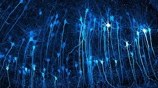
As imaging technology advances, so too does our understanding of cellular processes. Hickey’s video shows that mitochondria are more dynamic than previously thought, which suggests that they are “more than just a ‘powerhouse of the cell,’” he said. We’ve chosen to feature a several winners and honorable mentions that reveal the intricacies of the cellular world.
Extract — continue reading at the source.