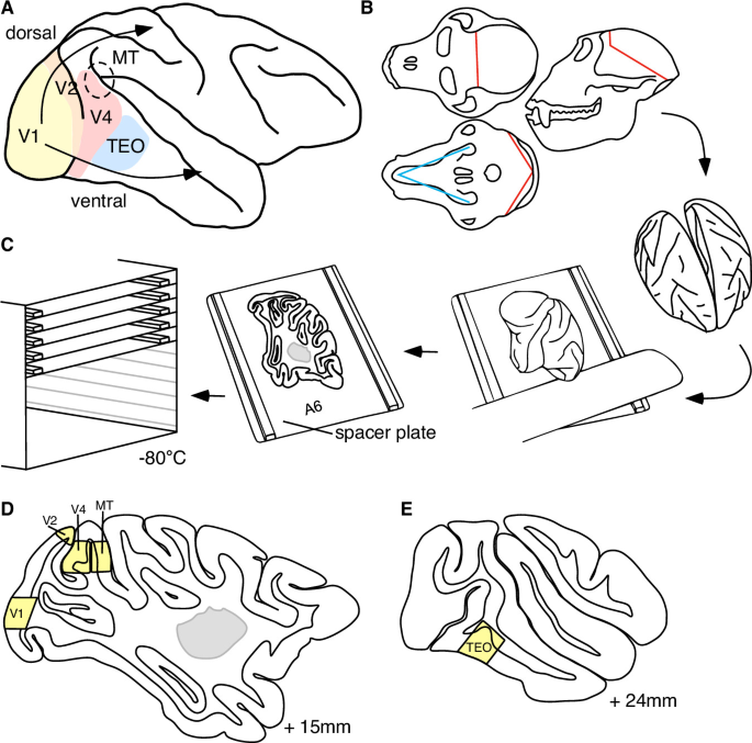
Non-human primates like rhesus macaques are pivotal models for decoding human visual cortex physiology and disorders. However, there is a lack of high-resolution transcriptomic atlases comparing distinct areas of the visual cortex in macaque. Here, we developed BrainSPACE, an innovative pipeline for rapid and precise brain tissue banking, to sample visual cortex areas: V1, V2, V4, MT, and TEO.
We applied single-nucleus RNA-sequencing (snRNA-seq) to unravel neuronal diversity in BrainSPACE-banked V1 and V4 areas ( ~ 100,000 nuclei). We uncovered conserved GABAergic neuron profiles but stark area-specific diversity in principal neurons, featuring seven unique layer 4 subtypes in V1 and one in V4. Complementary smFISH validated transcriptional gradients across these areas, aligning with ventral and dorsal stream hierarchies.
Gene ontology analyses highlighted plasticity-related pathways in unique layer 4 subtypes, with genes like NTNG1 and NLGN1 linked to neurodevelopmental disorders such as autism and schizophrenia. Our insights bridge molecular architecture to visual processing, offering an interactive atlas for community use. By revealing how layer 4 heterogeneity drives hierarchical specialization, our work advances primate brain mapping and informs therapeutic strategies for vision-related pathologies.
Non-human primates (NHPs) such as rhesus macaques are essential for understanding human brain physiology and disorders, particularly visual information processing. Studies in macaques have delineated the visual cortex into specialized areas, including the primary visual cortex (V1) and V4, a core model for higher visual processing. V1, also known as the striate cortex, featuring the prominent stria of Gennari, a band of heavily myelinated axons within layer 4 sublayers that is visible even to the naked eye in unfixed human brain tissue, sits at the bottom of the visual hierarchy, receiving thalamic input for initial processing1,2,3.
From there, information flows to higher areas via two streams: the ventral stream (V1 → V2 → V4 → TEO → TEav and TEpv) performing object recognition4, and the dorsal stream (MT → MST → LIPd → LIPv → PF/PFG) performing motion and spatial representation5. Thalamic input enters V1 through layer 4. Extensive research has delineated distinct sublayers in V1 layer 4 with differential connectivity patterns6, including a high proportion of spiny stellate cells7.
Feedforward projections from V1’s supragranular layers (2/3) target V4’s layer 43,8. Feedback from V4 to V1 originates primarily in infragranular layers (5/6)8 and avoids V1’s layer 43. Thus, V1 and V4 differ in signal reception, processing, and transmission layers.
Extract — continue reading at the source.


