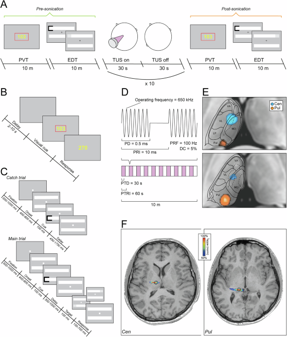
Using low-intensity focused transcranial ultrasound stimulation, we test the causal roles of the central thalamus in arousal and the pulvinar in visuospatial attention in the healthy human brain. In a within-subjects, counterbalanced design, 27 healthy volunteers (two females) complete a vigilance task (Psychomotor Vigilance Task) and a visuospatial attention task (Egly-Driver Task) before and after sham, central thalamic, and pulvinar sonication. Targeting the central thalamus slows responses and increases lapses in attention during the vigilance task, indicating reduced arousal.
Pulvinar sonication reduces (and slows) responses to visual stimuli presented contralaterally, suggesting a disruption of contralateral visuospatial attention. These results provide causal evidence linking the pulvinar to visuospatial attention and the central thalamus to arousal in the healthy human brain and demonstrate that ultrasound can elicit distinct behavioral changes in healthy volunteers when delivered to close but functionally distinct thalamic regions, underscoring its potential as a tool for noninvasive brain mapping. The thalamus and its interactions with the cortex play a central role in perception, attention, and consciousness.
However, because common neurostimulation techniques that are safe to use with healthy volunteers, such as transcranial magnetic stimulation (TMS), cannot directly target subcortical tissue, current understanding about the functional roles of deep brain structures like the thalamus in the healthy human brain relies on either correlational neuroimaging studies with healthy volunteers or lesion and invasive neurostimulation work in nonhuman animal models and human patient populations. The emerging technique of low-intensity focused transcranial ultrasound stimulation (TUS) addresses this gap by allowing safe, noninvasive modulation of subcortical tissue in the healthy human brain1. Using TUS, we aim to test the causal roles of two close but functionally distinct regions of the thalamus in the healthy human brain: the pulvinar and the central thalamus.
A growing body of work implicates the central thalamus, which comprises the intralaminar thalamic nuclei and adjacent tissue, in arousal regulation and the level of consciousness. This region not only bridges the brainstem and cortex along the ascending reticular activating system2 but also, through its extensive reciprocal connections with the cortex, is well positioned to facilitate cortical synchrony and electrocortical arousal3. Indeed, invasive electrical stimulation of the central thalamus in awake nonhuman primates can increase signs of arousal4 or induce lapses in consciousness5, depending on the stimulation parameters.
Strikingly, behavioral and electrocortical markers of arousal have been restored by targeting the central thalamus with pharmacological activation in anesthetized rodents6,7,8 and invasive electrical stimulation in anesthetized nonhuman primates9,10,11,12. Moreover, invasive deep brain stimulation and, more recently, TUS to the central thalamus in patients with disorders of consciousness (DOC) can yield improvements in their level of consciousness13. The pulvinar nucleus, which subsumes the posterior third of the thalamus, shows activation and interactions with the cortex that are consistent with a role in visual processing and attention.
The pulvinar is reciprocally connected with both visual cortices and higher-order frontal and parietal areas14. Visual responses modulated by attention can be recorded from the pulvinar15 and greater pulvinar activation is observed under states of engaged visual attention16. Beyond showing attention-dependent activation, the pulvinar also appears to play an active, coordinating role in visuospatial attention.
Extract — continue reading at the source.


