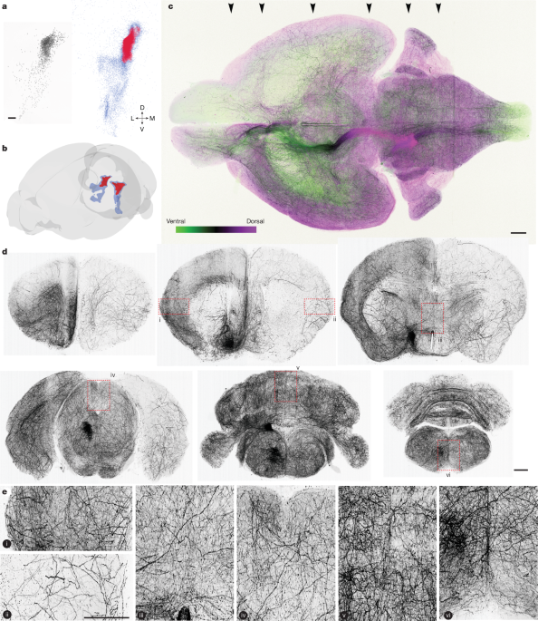
Noradrenaline (also known as norepinephrine) is released throughout most of the central nervous system by neurons in the locus coeruleus1,2,3,4. Here we identified a relationship between the morphologies, gene expression and activity of noradrenergic neurons in locus coeruleus in mice. Axonal projections of individual neurons were extensive but largely confined to subsets of brain regions.
Axonal projections and graded gene expression correlated with locations of cell bodies in locus coeruleus. In a behavioural task requiring continuous learning from actions, dorsal locus coeruleus neurons projecting to isocortex were activated when mice switched choices and by reward-prediction-error signals that drive learning. Background activity of neurons in ventral locus coeruleus was higher when mice ignored stimuli indicating potential reward availability.
These observations reveal a topographically organized structure and function of a neurotransmitter system and show that it contains learning signals for flexible behaviour. Monoamine neurotransmitters are released by small populations of neurons that project extensively throughout the central nervous system (CNS). The locus coeruleus (LC) in the pons supplies the monoamine noradrenaline to most of the CNS1,2,3,4.
Noradrenaline binds G-protein-coupled receptors on neurons and glia, activating intracellular signalling pathways5 and gating synaptic plasticity6,7. Noradrenaline release has diverse effects on cellular activity across brain targets8,9,10. Noradrenergic neurons in the locus coeruleus (LC-NE neurons) regulate multiple aspects of behaviour, including arousal11,12, sleep–wake transitions13, stress responses14,15, attention16,17 and learning18,19,20.
The diversity of functions of LC-NE neurons is often ascribed to their complex axonal projections, which span most of the brain and spinal cord. Although the LC-NE system has often been treated as homogeneous, several observations point to heterogeneity among LC-NE neurons21. LC neurons with different somatic morphologies show biases in their axonal projections22.
Ex vivo23,24,25 and in vivo26 electrophysiological recordings show diverse biophysical features in LC neurons. Subpopulations of LC-NE neurons exhibit distinct input–output patterns27 and have different functions in different CNS regions28,29. Heterogeneity among LC-NE neurons could thus underlie their diverse functions.
Extract — continue reading at the source.


