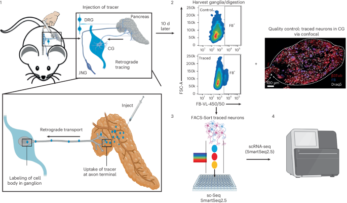
Neuronal innervation plays a pivotal role in controlling tissue function during homeostasis and regeneration, as well as in pathological contexts such as inflammation, autoimmune disorders, fibrosis and cancer. A major obstacle in understanding neuron–tissue interactions at the molecular level is that neuronal cell bodies reside in peripheral ganglia often far outside the organ of interest and are therefore typically excluded in common single-cell datasets of tissues. Here we developed Trace-n-Seq to address this issue—a method that combines retrograde tracing with fluorescence-activated cell sorting and single-cell sequencing to molecularly profile individual neurons innervating healthy or diseased tissues.
Trace-n-Seq can be used to explore the specific gene expression signatures of peripheral neurons at single-cell resolution. The method utilizes retrograde axonal tracing using Fast Blue, ganglia dissociation and fluorescence-activated cell sorting followed by single-cell RNA sequencing to enable the isolation and transcriptomic analysis of ganglia. Unlike bulk or whole-ganglia single-cell RNA sequencing without retrograde labeling, Trace-n-Seq allows the characterization of organ-specific neurons with single-cell resolution, eliminating background from uninvolved neurons and unrelated cell types located in the same ganglion.
The procedure allows researchers to obtain molecular transcriptomes of tissue-innervating neurons and to study their plasticity, subtype identity and potential interactions with the tissue microenvironment. The complete Trace-n-Seq workflow can be performed in 2 weeks and is suitable for researchers with experience in molecular biology, neuronal tissue handling, sequencing techniques and bioinformatic analysis. Trace-n-Seq integrates retrograde fluorescent tracing, FACS-based neuron isolation and single-cell RNA sequencing to profile ganglia complexity, including subsets of neurons that innervate specific tissues.
The method is versatile and applicable to various healthy, inflamed or diseased tissues innervated by the peripheral nervous system. This Protocol offers several advantages over existing approaches, including the ability to selectively profile neurons on the basis of functional tissue connectivity, capture rare tissue-specific neuronal subsets, distinguish disease-associated neuronal states from bulk ganglia signatures and directly compare neuronal remodeling across physiological and pathological conditions at single-cell resolution. This is a preview of subscription content, access via your institution Access Nature and 54 other Nature Portfolio journals Get Nature+, our best-value online-access subscription Receive 12 print issues and online access Prices may be subject to local taxes which are calculated during checkout All data supporting this Protocol are available within the Article and its Supplementary Information.
Additional raw and processed single-cell data can be provided upon reasonable request to the corresponding authors. The sequencing data are available via E-MTAB-12940. A repository containing the complete SMART-seq2 Trace n Seq analysis workflow, quality control scripts, reference-based neuronal annotation, Seurat pipelines and code to generate figures for healthy, pancreatitis and cancer neuron datasets can be found via GitHub at https://github.com/ManuelMastel/Trace-n-Seq.
Cancer research needs neuroscience and neuroscientists. Neurosci. 28, 915–917 (2025). Neuroanatomy Through Clinical Cases (Sinauer Associates, 2010).
Extract — continue reading at the source.


