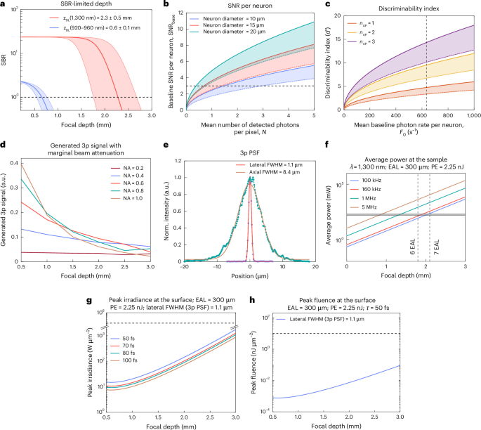
Extending the depth reach of multiphoton microscopy for imaging neuronal population activity in the scattering mammalian brain is an ‘exponentially hard’ problem. Three-photon (3p) microscopy has enabled imaging of neuroactivity at depths up to ~1.2 mm in the mouse brain; however, the fundamental depth limit of 3p imaging, as set by the depth at which out-of-focus background fluorescence exceeds the in-focus fluorescence signal, is well beyond 2 mm. Reaching this fundamental depth limit in 3p imaging has been hampered by insufficient in-focus fluorescence signal.
Here we demonstrate a three-photon ultra-deep imaging (TrUDI) system that approaches this fundamental depth limit via systematic and principled optimization of the in-focus fluorescence signal. TrUDI matches the parameters of the excitation laser, the optical system, and the acquisition to linear and nonlinear limits for safe laser exposure while satisfying the spatiotemporal requirements of single-cell resolution calcium (Ca2+) imaging. TrUDI achieves a ~250-fold increase in detected fluorescence photon yield at the same laser power as in previous approaches thereby enabling in vivo cellular-resolution recordings of neuronal population activity within fields of view of ~230 × 230 µm2 at 8.6 Hz at tissue depths of up to ~2 mm in mouse cortex and hippocampus.
This is a preview of subscription content, access via your institution Access Nature and 54 other Nature Portfolio journals Get Nature+, our best-value online-access subscription Receive 12 print issues and online access Prices may be subject to local taxes which are calculated during checkout The raw data are available from the corresponding author upon reasonable request as the data size is too large for sharing with public repositories. Source data are provided with this paper. Neuronal segmentations based on anatomical or spatiotemporal feature, and motion-corrections were implemented in MATLAB (R2024b) using open-source packages and are publicly available.
All custom codes used for data analysis in this work are provided as a compressed archive and are available with the paper as Supplementary Code 1 and can also be accessed from GitHub at https://github.com/vazirilab/TrUDI-ultradeep-3p, under an open-source license permitting not-for-profit research use (a License.md file is available with the code repository). Stringer, C., Pachitariu, M., Steinmetz, N., Carandini, M. & Harris, K. High-dimensional geometry of population responses in visual cortex.
Nature 571, 361–365 (2019). Article CAS PubMed PubMed Central Google Scholar Kafashan, M. et al. Scaling of sensory information in large neural populations shows signatures of information-limiting correlations.
Understanding sensory information processing through simultaneous multi-area population recordings. Neural Circuits 12, 115 (2019). Article PubMed PubMed Central Google Scholar Müller, E.
Extract — continue reading at the source.