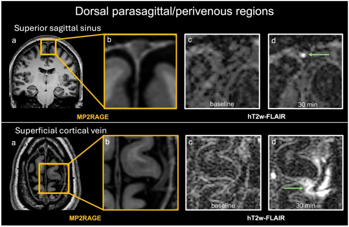
Brain fluid exchange can be studied in vivo with post-contrast MRI, but serial semiquantitative 7 T heavily T2-weighted FLAIR data in healthy adults remain limited. In this prospective study, we characterized post-contrast signal kinetics in CSF spaces and CSF outflow-related regions up to four hours after intravenous gadolinium administration. Fifteen healthy volunteers underwent 7 T MRI before and at 30, 90, 180, and 240 min after gadoterate meglumine administration.
Signal intensity (SI) was measured manually in predefined dorsal parasagittal/perivenous regions, ventral perineural regions, and CSF spaces on heavily T2-weighted 3D FLAIR. SI ratios were calculated relative to the splenium, and linear mixed models were used to test time-dependent change. A sensitivity analysis used the choroid glomus as alternative reference.
All sampled compartments showed significant time-dependent signal change. Dorsal parasagittal/perivenous regions showed rapid SI-ratio increase by 30 min with later decline, Meckel’s cave showed a similar but slower pattern, and the optic nerve sheath peaked later. CSF spaces showed smaller and slower changes.
The main temporal patterns in dorsal and ventral regions persisted with glomus normalization, whereas CSF-space changes were attenuated. These findings provide a normative 7 T semiquantitative benchmark and show distinct compartment-specific post-contrast temporal profiles together with inter-individual variability. Brain solute clearance is increasingly viewed as a distributed process involving exchange between blood, cerebrospinal fluid (CSF), interstitial fluid, perivascular spaces, and meningeal/dural compartments.
In rodents, periarterial CSF influx with subsequent perivenous efflux gave rise to the glymphatic concept, and subsequent work identified meningeal lymphatic vessels as an additional route linking cranial fluid drainage to the deep cervical lymph nodes1,2,3,4. At the same time, newer anatomical studies suggest that parasagittal dural channels and arachnoid cuff exit (ACE) points around bridging veins may also participate in CSF–dura communication, indicating that cranial outflow is unlikely to be confined to a single anatomical substrate5. In humans, MRI has become a major in vivo tool for studying brain fluid exchange.
Post-contrast MRI has demonstrated enhancement in parasagittal regions, along bridging-vein/perivenous pathways, around cranial nerve sheaths, and within selected CSF spaces5,6,7,8. However, after intravenous gadolinium administration, MRI signal in these regions cannot by itself distinguish lymphatic lumina from adjacent dural channels/stroma, or from communicating CSF-containing compartments, and should therefore be interpreted cautiously8,9,10. Several MRI approaches have been used for this purpose, including T1-weighted, T1-mapping, Fluid-Attenuated Inversion Recovery (FLAIR), and heavily T2-weighted FLAIR (hT2w-FLAIR) techniques7,11,12,13,14,15.
Extract — continue reading at the source.


