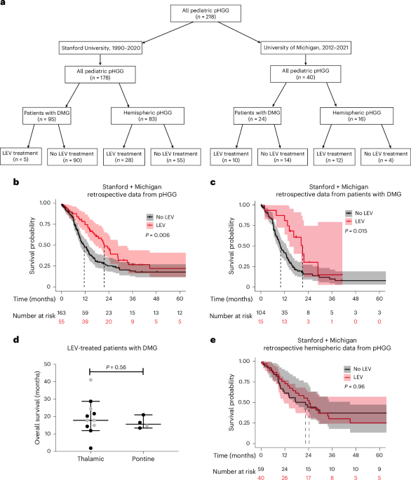
Antiseizure medications, such as levetiracetam, are commonly used in glioma patients. Retrospective analyses evaluating effects of antiseizure medications on glioma patient survival have yielded inconsistent results, probably due to confounding factors of glioma subtype and drug class. Here we present retrospective real-world clinical data that demonstrate longer overall survival for children with diffuse midline glioma (DMG) who were taking levetiracetam, which was not evident in pediatric patients with hemispheric high-grade glioma.
In preclinical models, levetiracetam reduces glioma proliferation and tumor burden, extending survival of mice bearing DMG orthotopic xenografts. These beneficial effects were not found in hemispheric high-grade glioma patient-derived orthotopic xenograft models. The subtype-specificity of this antiproliferative effect of levetiracetam is congruent with recent findings that GABAergic neuron-to-glioma synapses promote glioma growth in DMG but not hemispheric high-grade glioma.
We demonstrate here that levetiracetam attenuates low-frequency GABAergic synaptic transmission in a glioma-specific manner, reducing GABAergic synaptic currents in DMG but not in healthy neurons. This effect is independent of action on SV2A, the chief mechanism by which levetiracetam functions to prevent and treat seizures. Taken together, these findings indicate a promising approach to target GABAergic synapses in DMG and suggest that use of levetiracetam in DMG should be further studied in future prospective clinical studies.
High-grade gliomas (HGGs) are the leading cause of brain tumor-related morbidity and mortality in children. Pediatric-type gliomas are broadly classified1 as hemispheric HGGs that occur in the cerebral hemispheres and diffuse midline gliomas (DMGs) that occur in midline central nervous system structures, most commonly the brainstem, thalamus or spinal cord, and are often associated with H3K27M mutations in genes encoding histone H3. Neurons robustly drive the growth and progression of gliomas in a neuronal activity-regulated manner2 through both activity-regulated paracrine factor signaling2,3,4,5,6,7 and bona fide, electrophysiologically functional neuron-to-glioma synapses6,8,9,10,11.
Glioma cell membrane depolarization, through excitatory postsynaptic currents or other mechanisms, promotes glioma cell proliferation and tumor growth6,8 via voltage-sensitive mechanisms that remain to be fully elucidated. Just as neuronal activity promotes glioma growth and progression, gliomas increase neuronal excitability8,12,13,14,15,16. This glioma-induced neuronal hyperexcitability promotes glioma-associated seizures12,13,14,15 and further augments this pathogenic cycle of neuron–glioma interactions.
So far, glutamatergic neurons have been most extensively studied in this regard, and glutamatergic neuron-to-glioma synapses mediated by AMPA glutamate receptors are present in both DMG and hemispheric HGGs8,9. GABAergic neuron-to-glioma synapses are found in DMG where GABAergic synaptic signaling is depolarizing due to high intracellular chloride concentration in DMG malignant cells11. GABAergic neurons powerfully promote DMG growth, and medications that augment GABAergic signaling—for example, benzodiazepines such as lorazepam—accelerate DMG growth and shorten survival in patient-derived orthotopic xenograft models11.
Extract — continue reading at the source.


