Parallel processing, the ability to split the information content of sensory stimuli and handle them simultaneously, is essential for sensory perception in many systems. Mitral (MCs) and tufted cells (TCs) in the olfactory bulb (OB), with their separate but overlapping projection areas, provide an ideal anatomical basis for parallel processing. Whether they convey different information to distinct areas of the olfactory cortex remains unclear.
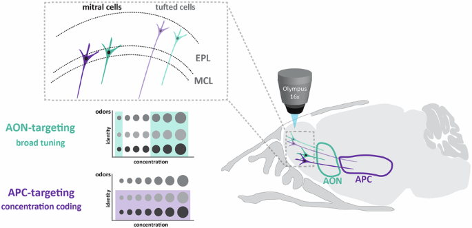
Here, we separated mouse OB output neurons by innervation area using retrograde tracing from the anterior olfactory nucleus (AON) or the anterior piriform cortex (APC). In vivo 2-photon calcium imaging in the OB revealed projection area-dependent differences, with APC-projecting neurons coding more strongly for odor concentration and more selectively for odor identity. This separation became more pronounced when differentially traced populations were split by cell type, with APC TCs displaying the strongest concentration-dependent changes in response and AON TCs showing very promiscuous odor tuning.
Purely cell-type-derived properties did not explain the projection target-dependent differences in odor representation, as both mitral and tufted cells showed similar changes, and SVM classifier analysis demonstrated that classification by projection target outperformed classification by cell type. Our data suggests a prominent projection-defined separation of sensory information in the OB. In a constantly changing environment, animals rely on rapid sensory uptake and efficient processing to respond adequately.
In the brain, the rapid processing of complex sensory stimuli is facilitated by parallel processing, which divides stimulus features into distinct channels, allowing computations to be performed simultaneously rather than sequentially. This principle is well established for the auditory, somatosensory, and visual systems1,2,3. Since chemical features are immensely complex, parallel processing has been postulated to be especially advantageous for the olfactory system4,5,6.
So far, only a few studies have investigated this in detail. The olfactory system is well-equipped for the parallel processing of sensory information. Olfactory bulb (OB) output neurons project to anatomically and functionally diverse olfactory cortical regions, including the olfactory tubercle (OT), anterior olfactory nucleus, piriform and lateral entorhinal cortex, and the amygdala.
While the cortical amygdala has been linked to innate, odor-driven behaviors7, the piriform cortex (PC) is implicated in odorant identity coding8,9, and olfactory memory10,11, the AON in storing episodic odor memories12,13, control of food intake14 and social odor processing15,16,17,18,19,20 and the OT is thought to be responsible for integrating sensory information and information about motivational states to guide affective and behavioral responses21. Accordingly, neuronal recordings from these different cortical areas show area-specific response properties, such as odor selectivity, integration of components in an odorant mixture, and experience-dependent plasticity20,22,23,24,25,26,27,28,29. While frequently treated as a single population of neurons, mitral (MCs) and tufted cells (TCs), the two principal OB output cells, target different areas in the olfactory cortex and are excellent candidates to initiate parallel processing in olfaction6,30,31,32,33.
Extract — continue reading at the source.


