The NLRP3 inflammasome has emerged as a critical regulator in neuroinflammatory responses to various brain insults from traumatic, ischemic and neurodegenerative conditions, an effect almost exclusively attributed to microglial NLRP3. Here, we demonstrated that in the absence of changes in inflammatory factors IL-18, IL-1β and TNF-α, neuronal Nlrp3 knockout in hippocampus (nNlrp3-/-) improved long-term spatial memory performance, with increased synaptic proteins levels and enhanced amplitude of long-term potentiation at 4 weeks after traumatic brain injury (TBI), compared to wildtype littermates. Notably, the nNlrp3-/- mice out-performed microglia-specific knockout and global Nlrp3 knockout mice in cognition and synaptic function.
We further identified that NLRP3 structurally and functionally directly interacted with the R1 subunit of N-methyl-D-aspartate receptor (NR1) in hippocampus after TBI. Accordingly, pharmacological inhibition of NLRP3 by MCC950 rescued the decrease in the NMDAR-gated calcium influx by LPS-stimulated hippocampal neurons while selective Nlrp3 knockout in hippocampal neurons partially reversed the decreased phosphorylation level of NR1 after TBI. Collectively, this study provides the first evidence that neuronal NLRP3 constitutes a non-inflammatory response to control synaptic plasticity and cognitive function via NR1-dependent non-inflammatory mechanism in TBI and thus broadens the central role of NLRP3 in neuronal adaptive mechanisms to include both microglial NLRP3-inflammation arm and the neuronal NLRP3-synaptic plasticity arm.
The persisting neuroinflammatory response is one of the most common characteristics of various brain insults from traumatic, ischemic and neurodegenerative conditions [1,2,3,4]. Increasing evidence revealed that the NLRP3 inflammasome is involved in many pathological processes of these diseases, and therefore contributes to cognitive impairment following these diseases, including traumatic brain injury (TBI) [5,6,7,8]. The proinflammatory effects of NLRP3 inflammasome is almost exclusively attributed to microglia in the most of these researches, and is considered a promising therapeutic target [9,10,11,12].
However, an increasing body of evidence has demonstrated that neurons themselves are also capable of expressing NLRP3 and its associated components [3, 13,14,15]. but the underlying molecular mechanisms of NLRP3 inflammasome from different types of neurocytes remain elusive. Another research has shown that following chronic constriction injury, NLRP3 inflammasome components are significantly upregulated in spinal cord neurons, and NLRP3 inflammasome activation via the NF-κB pathway promotes neuronal pyroptosis [15]. Studies on Parkinson’s disease (PD) also found the NLRP3 expression in dopamine neuron under pathological conditions, and the activation is associated with the neurodegenerative and necrosis of dopamine neurons via the regulation of NLRP3 phosphorylation and ubiquitination [3, 14].
So far, it has been proved that the NLRP3 inflammasome in microglia is involved in memory impairment via indirectly affecting neuronal function through its proinflammatory roles [16]. Inhibiting the activation of NLRP3 inflammasome in the acute (within 24 h) [17, 18] and chronic phases (over 2 months post-TBI) [19] can alleviate cognitive impairment and improve prognosis after TBI. These observations raise a key question: why does NLRP3 inflammasome inhibition remain therapeutically beneficial in the late phase of injury when proinflammatory cytokine levels are mildly elevated or have nearly fall back to baseline levels.
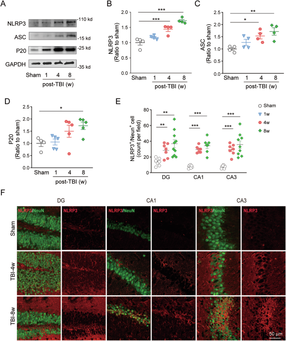
Although both inflammatory factors and pathological proteins can cause morphological and molecular alterations at the synapse, the changes of synaptic structure and electrical activity from neuron would have a more direct impact on the neuronal communication and neurobehaviors. Our previous study has found that the neuronal levels of NLRP3 increased after TBI [20], which prompt our consideration of the roles of neuronal NLRP3 that might be overlooked under TBI condition. In addition, published literature [21] and our studies [20, 22] have found that there exist impaired long-term potentiation (LTP) and a decline in hippocampus-related cognitive function in the chronic phase (4–8 weeks) of TBI.
Extract — continue reading at the source.