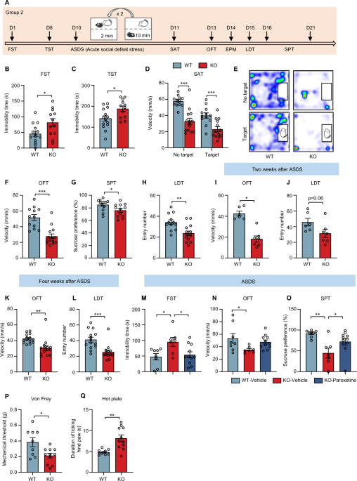
Neuroplasticity and synaptic dysfunction are central to the pathophysiology of depression, yet their underlying mechanisms remain not fully understood. Here, we identify the m6A reader protein YTHDF1 as a key regulator of depression pathogenesis. Mice with genetic deletion of Ythdf1 exhibited anterior cingulate cortex (ACC) hyperactivity, hyperalgesia, heightened social stress sensitivity, and anxiodepressive-like behaviors following acute social stress.
Using multi-omics approaches, we linked these effects to dysregulated actin cytoskeleton dynamics and identified cyclase-associated protein 1 (CAP1) as a downstream effector of YTHDF1. YTHDF1 deficiency reduced CAP1 expression from early postnatal stages, leading to F-actin accumulation, impaired synaptic function, and behavioral deficits. Strikingly, early postnatal re-expression of YTHDF1 in the ACC normalized CAP1 levels, restored synaptic integrity, and reversed depressive-like phenotypes.
Similarly, CAP1 re-expression was sufficient to alleviate stress-induced psychomotor retardation and anxiety. Our findings establish the YTHDF1-CAP1 axis as a critical pathway governing synaptic function and depressive behaviors. Major depressive disorder (MDD) has been recognized as one of the most concerning mental disorders worldwide due to its high prevalence, disabling effects, multiple comorbidity and complications [1, 2].
Despite extensive research, the complex pathogenesis of MDD remains poorly understood [3]. Accumulating evidence suggests that synaptic plasticity, which refers to the dynamic changes in synaptic structure and function, plays a crucial role in the pathological changes associated with mood disorders, especially depressive disorder [4, 5]. However, the intricate and dynamic process of synaptic plasticity is influenced by many factors.
Epigenetic mechanisms, such as DNA methylation and histone modifications, serve as mediators of the effect of environmental stressors on vulnerability-related genes [6], highlighting their pivotal roles in the onset and progression of MDD. Nonetheless, the impact of epitranscriptomics-mediated by RNA modifications on synaptic plasticity and depression remains largely unknown. As the most abundant internal modification in eukaryotic mRNA, N6-methyladenosine (m6A) is dynamically and reversibly regulated by methyltransferases (writers, such as METTL3/METTL14 complex) and demethylases (erasers, such as FTO and ALKBH5), which affects RNA splicing, trafficking, stability and translation through different readers [7]. m6A is highly expressed in the brain and localizes to synapses, contributing to various neuronal functions, including cortical neurogenesis [8], embryonic neural development [9], circadian clock [10], dopaminergic midbrain circuitry [11], memory formation and processing [12, 13], synaptic function [14], stress response regulation [15].
Previous studies have shown that the mRNA levels of m6A writers and erasers were altered in the peripheral blood and specific brain regions of patients with MDD as well as animal models of depression [16, 17]. For example, selective knockout of FTO in the hippocampus induces depressive-like behavior [16], while targeted deletion of ALKBH5 in astrocytes exerts antidepressant-like effects [17]. Additionally, neuropathic pain has been shown to reduce FTO expression in the anterior cingulate cortex (ACC), contributing to depressive-like behaviors [18].
Extract — continue reading at the source.


